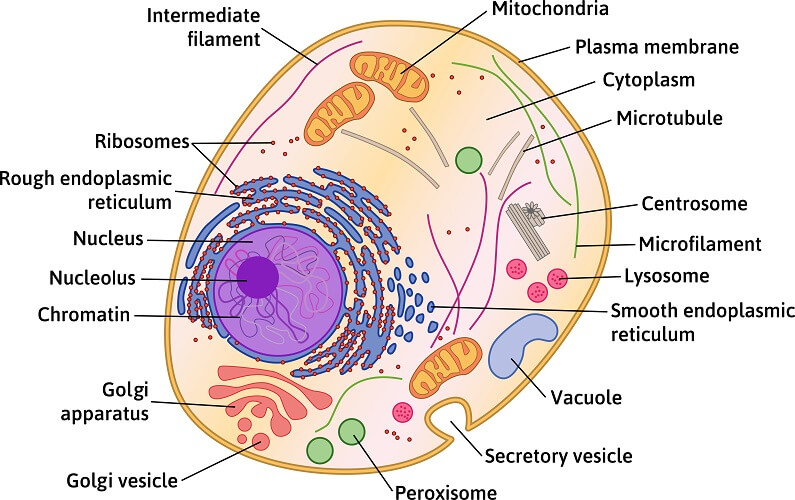
Animal Cell Diagram Microtubules / Animal Cell National Geographic Society : Smooth endoplasmic reticulum, mitochondria, golgi bodies, lysosomes.
byZackary Gambale-0
Animal Cell Diagram Microtubules / Animal Cell National Geographic Society : Smooth endoplasmic reticulum, mitochondria, golgi bodies, lysosomes.. Microtubules can grow as long as 50 micrometres and are highly dynamic. Microtubules as their name implies, microtubules are small hollow tubes. Their primary function is to help support and shape the cell. In the complete animal cell centrosome, the two centrioles are arranged such that one is perpendicular to. This note contains detail information about microbodies microtubules peroxisomes are the microbodies found in many animal cells and in a wide range of plants.
The cellular organization of microtubules varies between cell types, but in most cells, the minus ends of microtubules are anchored to the centrosomes near the nucleus while the plus ends radiate towards the periphery of the cell. A cell carries out all the processes of the body which includes producing energy and storing it, making proteins • microtubules: In animal cells, microtubules radiate outwards from an organelle in the center of the cell called a centrosome, which is a microtubule organizing center (mtoc). All organisms are made up of cells (or in some cases, a single cell). Cell membrane is made up of lipids and proteins and forms a barrier between the extracellular liquid.
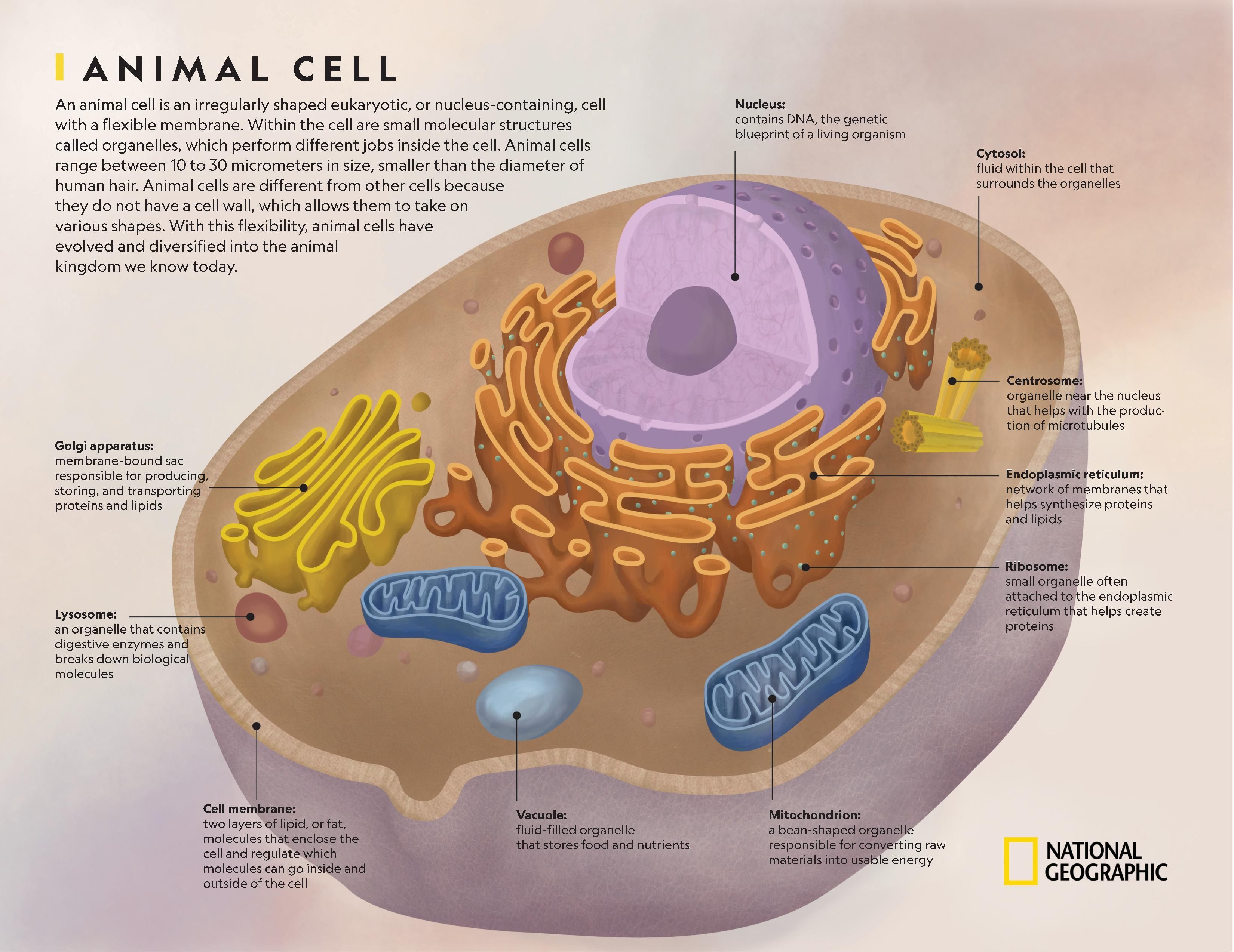
Animal Cell The Definitive Guide Biology Dictionary from biologydictionary.net The cells of plants and fungi do not have centrosomes, and instead the nuclear envelope—the membrane surrounding the cell's. Conduit pt et al., centrosome function and assembly in animal cells. Cell membrane is made up of lipids and proteins and forms a barrier between the extracellular liquid. Animal cells have a variety of different organelles that work together to allow the cell to perform its functions. Animal cell structures, functions & diagrams. The nucleus of a cell contains its dna and controls the cell's growth and. The role and function of the plasma membrane; In truth, there are still features of plant and animal cells we're only lately.
Microfilaments, microtubules, and intermediate filaments.
The cell is engaged in metabolic activity and performing its prepare for mitosis (the next four phases that lead up to and include nuclear division). An animal cell diagram is a great way to learn and understand the many functions of an animal cell. A comparison of plant and animal cells using labelled diagrams and descriptive explanations. Microtubules are the fibrous, hollow rods typically found in eukaryotic cells. They are stiff enough to prevent the cell from. They are about 23 nm in size which is the largest of the cytoskeleton fibers. These are present in all photosynthetic cells of higher. Our body starts its existence at fertilization from a single cell, the diploid cilia, flagella and centrioles are composed of microtubules. It is made up of 9 microtubule bundles and their primary function is to assist in organizing. Microtubule length is quite variable. Microtubules are polymers of tubulin that form part of the cytoskeleton and provide structure and shape to eukaryotic cells. Smooth endoplasmic reticulum, mitochondria, golgi bodies, lysosomes. Here is a summary of their structure and function.
They are about 23 nm in size which is the largest of the cytoskeleton fibers. Most animal cells are diploid, meaning that their chromosomes exist in homologous pairs. All organisms are made up of cells (or in some cases, a single cell). Microtubule length is quite variable. These are present in all photosynthetic cells of higher.
Animal Cell National Geographic Society from media.nationalgeographic.org Microtubules can grow as long as 50 micrometres and are highly dynamic. Both of these structures are found in animal cells, but not plant cells. Animal cells are the types of cells that make up most of the tissue cells in animals. The most important structures of plant and animal cells are shown in the diagrams below, which provide a clear illustration of how much these cells have in common. Conduit pt et al., centrosome function and assembly in animal cells. These are present in all photosynthetic cells of higher. Microtubule length is quite variable. The cells of plants and fungi do not have centrosomes, and instead the nuclear envelope—the membrane surrounding the cell's.
Microtubules can grow as long as 50 micrometres and are highly dynamic.
It is made up of 9 microtubule bundles and their primary function is to assist in organizing. Different kinds of animals have microtubules: That cells can be of different shapes and sizes. These are present in all photosynthetic cells of higher. The cell may contain a pair of centrioles (or microtubule organizing centers in plants) both of which are organizational sites for microtubules. Smooth endoplasmic reticulum, mitochondria, golgi bodies, lysosomes. Animal cells are the types of cells that make up most of the tissue cells in animals. Microtubules in the cell consist of microscopic structures formed in hollow tubes and constructed in a series of linear rings. Animal cells have a variety of different organelles that work together to allow the cell to perform its functions. Mitochondria since mitochondria provide the cell with energy, you as the diagram indicates, most of the microtubules radiate out from a central region within the cytoplasm. The role and function of the plasma membrane; But at the same time it is interpretive. A comparison of plant and animal cells using labelled diagrams and descriptive explanations.
Microbodies microtubules and microfilament are the additional cell organelles. Centrioles are bundles of cylindrical microtubules that separate the 2 cells. Each centriole is a ring of nine groups of fused microtubules. Different kinds of animals have microtubules: Microtubules can grow as long as 50 micrometres and are highly dynamic.
Animal Cell Diagram In Detail from www.anatomynote.com They are about 23 nm in size which is the largest of the cytoskeleton fibers. They are stiff enough to prevent the cell from. The walls of the microtubule are made of with a diameter of about 25 nm, microtubules are the widest components of the cytoskeleton. Centrioles are composed of groupings of microtubules arranged in a 9 + 3 pattern. Mitochondria since mitochondria provide the cell with energy, you as the diagram indicates, most of the microtubules radiate out from a central region within the cytoplasm. The cell is the basic unit of life. Microtubules can also be distinguished from microfilaments chemically. Different kinds of animals have microtubules:
Our body starts its existence at fertilization from a single cell, the diploid cilia, flagella and centrioles are composed of microtubules.
The outer diameter of a microtubule is between 23 and 27 nm while the inner diameter is between 11. Microtubules in the cell consist of microscopic structures formed in hollow tubes and constructed in a series of linear rings. Learn vocabulary, terms and more with flashcards, games and other study tools. Microtubules are polymers of tubulin that form part of the cytoskeleton and provide structure and shape to eukaryotic cells. Both of these structures are found in animal cells, but not plant cells. These are present in all photosynthetic cells of higher. Start studying animal cell diagram. The walls of the microtubule are made of with a diameter of about 25 nm, microtubules are the widest components of the cytoskeleton. Some microtubules are less than 200 nm long, but in the long processes of nerve cells their lengths may be as great as 25 μm (i.e., 25,000 nm). Here is a summary of their structure and function. Microtubules as their name implies, microtubules are small hollow tubes. During cell division, spindle formation takes place through microtubules in plant cells,but in animal cells it takes place through aster formation. Microtubules can also be distinguished from microfilaments chemically.


Post a Comment